The following points highlight the nine main methods and techniques of molecular biology. The methods are:- 1. DNA Ligation 2. Elution of DNA Fragments from Agarose 3. Phenol Purification of DNA from Low Melting Agarose 4. Polymerase Chain Reaction 5. SDS-Polyacrylamide Gel Electrophoresis 6. Iso-Electric Focusing (IEF) of Proteins 7. Trypsin Digestion of Protein Gel 8. Protein Dialysis 9. Enzyme (Esterase) Gel Electrophoresis.
Method and Technique # 1. DNA Ligation:
In order to construct new DNA molecules, DNA must first be digested using restriction endonucleases. The individual components of the desired DNA molecules are then purified and combined and treated with DNA ligase.
The term recombinant DNA encapsulates the concept of recombining fragments of DNA from different sources into a new, useful DNA molecule. Joining linear DNA fragments with covalent bonds is called ligation. More specifically DNA ligation involves creating a phosphodiester bond between the 3′ hydroxyl of one nucleotide and the 5′ phosphate of another nucleotide.
The enzyme used to ligate DNA fragments is T4 DNA ligase, which originates from the T4 bacteriophage. This enzyme will ligate DNA fragments having overhanging, cohesive ends (sticky ends) that are annealed together.
The products of ligation mixture are introduced into competent E. coli cells and transformants are identified using appropriate genetic marker selection.
1. Add the following to a microfuge tube –
10X Ligase buffer – 2 μl (1X final concentration)
Component DNAs – 2 μl (0.1 to 5 μg)
(Normally a mixture of vector and insert DNA)
10 mM ATP – 1 μl
T4 DNA ligase – 1μl (20 – 500 units)
Distilled water – 14 μl
2. Incubate for 1-24 hours at 15°C.
3. Keep in a refrigerator for 30 minutes.
4. Subject them to heat shock at 42°C for 2 minutes.
(Introduce 1-10 μl of the ligated products into competent cells and select for transformants using genetic marker present on the vector).
Method and Technique # 2. Elution of DNA Fragments from Agarose:
DNA fragments are eluted from low-melting temperature agarose gels. Here, the band of interest is excised with a sterile razor blade, placed in a micro-centrifuge tube, frozen at -70°C, and then melted.
Then, TE-saturated phenol is added to the melted gel slice, and the mixture is again frozen and thawed. After this second thawing, the tube is centrifuged and the aqueous layer removed to a new tube. Residual phenol is removed with two ether extractions, and the DNA is concentrated by ethanol precipitation.
1. Place excised DNA-containing agarose gel slice in a 1.5 ml micro-centrifuge tube and freeze at -70°C for at least 15 minutes, or until frozen.
(It is possible to pause at this stage in the elution procedure and leave the gel slice frozen at -70°C.)
2. Melt the slice by incubating the tube at 65°C.
3. Add one-volume of TE-saturated phenol, vortex for 30 seconds, and freeze the sample at -70°C for 15 minutes.
4. Thaw the sample, and centrifuge in a micro-centrifuge at 12,000 rpm for 5 minutes at room temperature to separate the phases.
5. Remove the aqueous phase to a clean tube, extract twice with equal volume of ether, precipitate using ethanol and rinse the DNA pellet with 70% ethanol, air dry and dissolve in TE or distilled water.
Method and Technique # 3. Phenol Purification of DNA from Low Melting Agarose:
1. Incubate the buffer saturated phenol at 37°C for 1 hour.
2. Melt excised agarose fragment at 65°C for 10 minutes or until agarose is molten, place molten agarose at 37°C for 2 minutes.
3. Add 0.5 volume of buffer saturated phenol and shake vigorously for 45 seconds.
4. Centrifuge at 10,000 rpm for 5 minutes at room temperature and transfer aqueous layer to a clean tube.
5. Repeat phenol extraction and centrifugation steps.
6. Transfer aqueous layer to a clean tube and add 1/50 volume of 5 M NaCl, mix thoroughly for 45 seconds and centrifuge at 10,000 rpm for 5 minutes at room temperature.
7. Transfer the aqueous material to a clean tube, add two volume of ethanol and precipitate the DNA by keeping at -70°C for 15 minutes or -20°C for 1 hour.
8. Centrifuge at 10,000 rpm for 15 minutes and pour off the aqueous phase.
9. Wash the pellet in 70% ethanol and repeat centrifugation.
10. Pour off the aqueous material and air-dry the DNA for 5 minutes or dry in Speedvac for 2 minutes.
11. Re-suspend the DNA in 20 μl TE buffer.
Method and Technique # 4. Polymerase Chain Reaction (PCR) (In Vitro Amplification of DNA):
Polymerase chain reaction (PCR) is a powerful tool that allows amplification of specific DNA sequences. Successful PCR includes the design of optimal primer pair, appropriate primer concentrations, optimization of PCR conditions.
Primers are most important for PCR reactions. The length of the primer ranges 18-30 nucleotides. The GC content also varies from 40% to 60%. The primers anneal to DNA templates at a known temperature and that is calculated based on the melting temperature (Tm) of the primer [Tm value = 2 (A+T) + 4 (G+C)].
Optimal Tm value is above or below the estimated Tm. It is usual to have annealing temperature 5°C below the Tin. Annealing temperature may also need to be optimized for efficient PCR.
Complementarities of two or three bases at the 3′ end of the primer pairs are avoided to reduce primer-dimer’ formation. To prevent mismatches between the 3′ end of the primer and the target-template sequence, runs of three or more G or C at the 3′ end are avoided. Primers with a T at the 3′ end have a greater tolerance of mismatch. Commercially available computer software can be used for primer design for known genes.
Use concentrations of 0.1 to 0.5 µM of each primer. For most applications, a primer concentration of 0.2 µM will be sufficient. The concentrations of primer and Mg2+ in the PCR buffer need to be optimized for each primer.
Normally the buffer delivered with the polymerase by one company is often not useable with enzymes from other manufacturers. NaCl concentrations >50 mM inhibit the Taq DNA polymerase.
MgCl2 must be added as Mg2+ is an essential cofactor for the DNA polymerase. The most common components of 10X buffers are – 500 mM KCl, 100 mM Tris-HCl pH 8.3, 1%-2% Trition X 100 or 0.1% Tween or 0.1% Gelatin and 10-15 mM MgCl2.
MgCl2 at 0.5 to 3.5 mM is essential as a co-factor of DNA polymerase in the assay buffer. If dUTP is used instead of dTTP the magnesium ion concentration usually must be increased (up to a maximum of 5 mM).
But higher concentration always promotes the amplification of unspecific fragments and also increases the melting temperature. Very low concentrations reduce annealing efficiency and the synthesis rate.
The dNTPs are usually stored in 10 mM concentrations at pH 7.0. A concentration of 20-200 µM is required for the PCR reaction. Higher concentration of dNTPs leads to mispriming and misincorporation of nucleotides. All nucleotides must have the same concentration.
Inclusion of control reactions (preferably a positive control without enzyme) is essential for monitoring the success of PCR reactions. A negative control (without template DNA) is also to be included to ensure that the solutions used for the PCR have not been contaminated with the DNA.
This DNA fragment contains three genes. Only gene 2 is to be amplified.
RAPD stands for random amplification of polymorphic DNA. RAPD reactions are PCR reactions that essentially amplify segments of DNA, which are unknown or random. PCR is very often used to amplify a known sequence of DNA.
In RAPD the target sequence is unknown. A primer with an arbitrary sequence is prepared for this. A ten base pair sequence is used as primer for most RAPD reactions.
Figure 4.2 depicts a RAPD reaction, where a large fragment of DNA is used as template (genomic DNA) in the PCR reaction containing many copies of a single arbitrary (random) primer.
i. The arrows represent multiple copies of the primer (all primers have the same sequence). The direction of the arrow also indicates the direction in which DNA synthesis will occur.
ii. The numbers represent locations on the DNA template to which the primers anneal.
iii. Primers anneal to sites 1, 2 and 3 on the top strand of the DNA template and to sites 4, 5 and 6 on the bottom strand of the DNA template.
In this example, only 2 RAPD-PCR products are formed.
1. Product A is produced by PCR amplification of the DNA sequence which lies in between the primers bound at positions 2 and 5.
2. Product B is produced by PCR amplification of the DNA sequence which lies in between the primers bound at positions 3 and 6.
Note that –
i. No PCR product is produced by the primers bound at positions 1 and 4 because these primers are too far apart to allow completion of the PCR reaction.
ii. No PCR products are produced by the primers bound at positions 4 and 2 or positions 5 and 3 because these primer pairs are not oriented towards each other.
If another DNA template (genome) was obtained from a different source, there would probably be some differences in the DNA sequence of the template.
The process of DNA synthesis can occur in only one direction (shown by the arrows). Thus, two primers must anneal to the template DNA on opposite strands, and in opposite orientations such that they face each other, in order to achieve synthesis of both strands of the DNA segment which lies between the two primers.
In the figure above, primers 2 and 5 do not face each other.
Primers 3 and 6 anneal to opposite strands and face each other, leading to the synthesis of product B.
Likewise there will be no amplification of the DNA which lies between primers 3 and 5.
In order for a PCR reaction to work, the primers must be within reasonable distance of each other.
Consider the reaction shown in the following figure (a typical PCR amplification reaction).
Specific Amplification of DNA:
Protocol:
Use a clean work surface for all PCR preparations. PCR mix can be prepared preferably in a laminar air flow chamber.
1. Prepare the PCR mix as follows (50 µL reaction volume)-
2. Mix well and centrifuge briefly.
3. Prepare a control reaction with no template DNA (negative control) and without enzyme (positive control).
4. Place the tubes in a thermal cycler preheated at 94°C.
5. Run the programme as follows-
(Use appropriate annealing temperature for a particular primer.)
6. Add 2.5 µl sample buffer after amplification in each amplified sample and check the amplified products using appropriate concentration of agarose gel followed by electrophoresis or store at 4°C.
Random Amplification of Polymorphic DNA (RAPD – PCR):
1. Prepare the PCR mix as follows (20 µl reaction volume) –
2. Mix well and centrifuge briefly.
3. Prepare a control reaction with no template DNA (negative control) and without enzyme (positive control).
4. Place the tubes in a thermal cycler preheated at 94°C and set the following cycling conditions –
5. Add 2.5 µl sample buffer after amplification in each amplified sample and check the amplified products using appropriate concentration of agarose gel followed by electrophoresis or store at 4°C.
Reverse Transcriptase-Polymerase Chain Reaction (RT-PCR):
RNA must be reverse transcribed [using a reverse transcription (RT) reaction] into cDNA for polymerase chain reaction using RNA as a template. RT and PCR can be carried out separately (one step RT-PCR) or in a single tube (two step RT-PCR). The one step RT-PCR requires gene specific primers.
Primers for RT-PCR should be designed in such a way that they should not amplify any contaminating DNA present in the RNA sample. This results when one half of the primer hybridizes to the 3′-end of one exon and the other half to the 5′-end of the adjacent exon as shown in Figs 4.6(a) and 4.6(b).
Such primers will anneal to the cDNA synthesized from the spliced mRNAs, but not to the genomic DNA. If only mRNA sequence is known, choose primer-annealing sites that are at least 300-400 bp apart. It is likely that fragments of this size from eukaryotic DNA contain splice junctions. Such primers may be used to detect DNA contamination.
Protocol:
Reverse Transcription (RT) Reaction:
1. Mix the following reagents in a microfuge tube –
2. Incubate at 37°C for 60 minutes.
(or refer to suppliers’ instructions for specific enzymes)
RT-PCR Reaction:
3. Use an aliquot (4 or 5 µl) of completed reverse transcription reaction to the PCR mix.
4. Carry out the PCR reaction.
Bacterial Colony PCR:
This protocol allows rapid detection of transformation success and determination of correct ligation products by size (used to determine whether or not a specific colony on a plate has a desired sequence).
Protocol:
1. Use primers specific for vector sequence for preparing the PCR reaction mix.
2. Select colonies and number on the lower side of the plate.
3. Prepare the PCR mix (at least for 15 colonies) for colony PCR as follows –
4. Add the mix to the PCR tubes (10 µl/reaction) on ice without adding the colony.
5. Using the pipette tip touch the selected colony on its side and stir the tip in the PCR mix. Carry out this for all the colonies to be analyzed.
6. Place the tubes in a thermal cycler and amplify the plasmid by following specific amplification protocol
Method and Technique # 5. SDS-Polyacrylamide Gel Electrophoresis:
This technique involves the separation of proteins based on their size. When heated under denaturing conditions, proteins become unfolded and coated with the anionic detergent sodium dodecyl sulfate (SDS), acquiring net negative charges irrespective of their intrinsic electrical charges.
When the sample is loaded on a gel, the negatively charged protein molecules migrate towards the positively charged electrode in an electric field. After gel electrophoresis, the size of the polypeptide can be estimated by comparing its migration distance with that of the standard molecular weight proteins.
The acrylamide gels are formed by polymerizing acrylamide with a cross-linker (bis-acryl- amide) in the presence of a catalyst (TEMED) and an initiator (ammonium persulfate) with a suitable gel buffer (Tris).
Solutions are normally degassed prior to polymerization. Oxygen molecules inhibit polymerization and additionally heat will be produced during polymerization. The rate at which the gels polymerize can be controlled by varying the concentrations of TEMED and ammonium persulfate.
Protein molecules can be separated by electrophoresis on the basis of –
Charge – using isoelectric focusing
Size – SDS-polyacrylamide gel electrophoresis (denatured gel electrophoresis)
Shape – Native polyacrylamide gel electrophoresis (non-denatured gel electrophoresis)
The most widely used system of gel electrophoresis is the discontinuous gel electrophoresis. In this system the gel is composed of ¾ of separating gel and ¼ of stacking gel with two buffer systems for making the gel. If the samples are loaded directly on the top of the gel the sharpness is lost and the protein band in the gel will be as broad as possible.
This problem is overcome by polymerizing a stacking gel on the top of the resolving gel. When electrophoresis started in such a system the proteins and ions migrate into the stacking gel. The proteins concentrate in a very thin zone called the stack between the fast moving Tris ion and the slow moving glycine ion. As a result the bands produced are very sharp and clear.
Protocol:
1. Thoroughly clean the plates with SDS (1%) and wash with water and then dry the plates.
2. Assemble the gel plates according to the manufacturer’s instruction and determine the volume of the gel mould.
3. Mark the level to which the separating gel mix should be poured, a few millimeters below the level where wells will be formed by the comb.
4. In a conical flask prepare the acrylamide separating gel mixture. Refer to the table for known percentage and volume of mix.
5. Mix the components and without delay pour the acrylamide mixture into the glass mould and overlay with distilled water or isopropylalcohol.
6. After polymerization (approximately 20-30 minutes) remove the overlay and wash the top of the gel with distilled water.
7. Prepare the acrylamide stacking gel mixture. Refer to the table for known percentage and volume of mix.
8. Mix the components and pour over the separating gel.
9. Immediately insert the comb and allow the gel to polymerize (approximately 10 minutes).
10. Place the gel mould in the electrophoresis chamber. Fill the chamber with electrophoresis buffer and remove the comb.
11. Prepare the samples (normally 100 µg protein) with equal volume of sample buffer (if sample volume is 10 µl add 10 µl sample buffer). Mix thoroughly.
12. Heat the samples in boiling water bath approximately for 2 minutes to denature the proteins. Reduce the heating time according to the quantity of the protein or volume of the sample. Do not over-cook the proteins. Keep them on ice to retain the denatured stage.
13. Load the samples and apply the current 8 volt/cm for stacking gel (70 volts) and 15 volt/cm for separating gel (150-200 volts) depending upon the dimension of the gel (mini or regular).
14. Run the gel until the bromophenol blue dye reaches the bottom of the resolving gel.
15. Turn off the power supply, remove the gel mould from the apparatus, and separate the plates using a spatula. Mark the orientation in the gel.
16. Immerse the gel in a tray containing 5 volumes of staining solution (normal staining is carried out with coomassie brilliant blue). Stain the gel at room temperature for 4 hours.
17. Remove the stain and destain the gel.
18. Store the gel in 7% acetic acid. Visualize the bands under an illuminator.
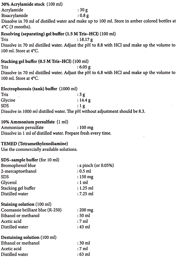
Preparation of Solutions:
Note:
The acrylamide forms the basic matrix in the gel. Monomer of acrylamide is a neurotoxin. Bis-acrylamide is a cross linker used to produce cross-linked matrix with a particular pore size. It is also a neurotoxin. When they polymerize they become non-toxic. Both have water releasing property. They approximately release 0.9 ml of water per gram.
Mercaptoethanol is essential in the sample buffer to ensure breakage of disulphide bonds when the sample is boiled.
Glycerol is used to increase the density of the sample. When sample is applied to the gel it remains as a well-defined overlay and does not undergo convective mixing with the electrophoresis buffer.
Sodium dodecyl sulfate is helpful in denaturing the protein samples. It provides a net negative charge to the cleaved proteins and thus separates them according to their molecular weight or size in this system. As all the polypeptides are negatively charged they migrate from cathode to anode in the gel.
Silver Staining of Proteins:
Visualization of the protein bands is carried out by incubating the gel either with coomassie brilliant blue R-250 or with silver staining. The coomassie stain can detect a band containing as little as 100 ng of protein. Such sensitivity is generally sufficient.
However, there are cases when greater sensitivity would be an advantage and this can be achieved by using silver stain. Silver staining is more sensitive than coomassie, and is able to detect 2-5 ng of protein. The sensitivity of silver stain is comparable to autoradiography of radio-labelled proteins.
During silver staining all glassware should be thoroughly cleaned and all solutions should be filtered. Gloves should be worn at all times. After use all silver nitrate solutions should not be flushed down the sink but added to HCl to precipitate silver chloride and destroy potentially explosive silver complexes.
Selective reduction of silver from ionic to metallic is the basic principle for the method. Sodium thiosulfate and formaldehyde are used as reducing agents. Addition of sodium carbonate to the gel which was soaked in the silver nitrate, ensure that the silver cations are complexes with the carboxyl group of proteins.
Protocol:
1. Fix the gel in 50% ethanol or methanol (50 ml), 12% acetic acid (12 ml) and 0.05% formaldehyde (50 µl) in distilled water (38 ml) for two hours to overnight.
2. Wash the gel with 50% ethanol or methanol (50 ml ethanol and 50 ml distilled water) three times each 20 seconds.
3. Treat the gel with Na2S2O3 (sodium thiosulfate, 20 mg dissolved in 100 ml) for 1 minute.
4. Wash the gel with distilled water three times each for 20 seconds.
5. Treat the gel with silver nitrate (400 mg in 200 ml distilled water with 160 µl formaldehyde) for 20 minutes.
6. Wash the gel with distilled water three times each for 20 seconds.
7. Develop the gel in 200 ml distilled water containing 6 g sodium carbonate, pinch of sodium thiosulfate and 50 µl formaldehyde.
8. Remove the developer and arrest the colour development by adding distilled water.
9. Preserve the gel in 12% acetic acid.
Extraction of Proteins:
Plant Material:
1. Homogenize the material (about 2 g) thoroughly in Tris-HCl buffer pH 7.2 (about 8-12 ml) using sterile glass powder in an ice bath.
2. Centrifuge the homogenate at 10,000 rpm for 10-15 minutes at 4°C.
3. Collect the supernatant.
4. Precipitate the proteins using 10% TCA (150-200 µl).
5. Keep it at -20°C for one hour.
6. Collect the protein precipitate by centrifugation at 10,000 rpm for 10 minutes at 4°C.
7. Wash the precipitate once with 5 ml of 80% acetone to extract residual TCA.
8. Repeat the centrifugation step.
9. Dissolve the pellet in 500 µl of Tris-HCl, pH 7.2.
Fungus:
1. Homogenize the mycelial mat in a pre-chilled mortar and pestle using cold 100 mM Tris-HCl buffer pH 7.0 containing 20 % sucrose, 0.1% cysteine hydrochloride and 0.1% ascorbic acid.
2. Centrifuge the homogenate at 10,000 rpm for 10 minutes at 4°C.
3. Collect the supernatant.
Seeds:
1. Remove the seed coat and make a fine meal of the seeds.
2. Grind 100 mg of seed powder in 1 ml of 100 mM Tris-HCl, pH 8.0 on ice.
3. Centrifuge at 10,000 rpm for 10 minutes at 4°C.
4. Collect the supernatant.
Animal Tissue:
1. Grind 100 mg of the tissue in 1 ml of 50 mM phosphate buffered saline pH 7.0 using a glass homogenizer.
2. Centrifuge at 10,000 rpm for 10 minutes at 4°C.
3. Collect the supernatant.
Preparation of Solutions:
Phosphate Buffered Saline, 50 mM (PBS):
50 mM NaH2PO4 – 780 mg
50 mM Na2HPO4 – 710mg
Dissolve in 80 ml distilled water.
Adjust the pH to 7.0.
Add 0.877 g NaCl (0.15 M) and 0.174 g PMSF (1 mM).
Make up the volume to 100 ml.
Bradford Protein Assay:
The Bradford protein assay is based on an absorbance shift in the dye Coomassie (G-250 grade) when bound to arginine and aromatic amino acid residues present in the protein. The anionic (bound) form has an absorption spectrum maximum at 595 nm whereas the cationic (unbound) form has an absorbance maximum at 470 nm.
The increase of absorbance at 595 nm is proportional to the amount of bound dye and thus, to the amount (concentration) of protein present in the sample. Unlike other protein assays, the Bradford Protein Assay is less susceptible to interference by various chemicals that may be present in protein samples.
Protocol:
1. Dilute Bradford dye concentrate with distilled water (see bottle for specifics; however 1ml dye + 4 ml water is common). Filter the dye solution (gravity filtration)—the diluted reagent may be used for approximately 2 weeks. A standard curve is needed to determine the concentration of the unknown samples.
2. In order to determine the protein concentration in the samples, you need to create a standard curve –
a. Using 6 cuvettes (1ml capacity), add the following amounts of BSA: 0 (baseline), 2, 5, 8, 10, 15 micrograms
b. Fill with Bradford dye (to a volume of 1 ml)
c. Carefully vortex the cuvettes and allow them to incubate for approximately 5 minutes at room temperature. Absorbance will increase with time, therefore be consistent with the amount of time that the samples are allowed to incubate before spectrophotometric measurement.
d. Using a spectrophotometer, measure the OD at 595 nm
3. Measure the OD of unknown samples; measure the OD of the extraction buffer (blank) to remove its contribution to total protein.
4. Plot the results for the BSA and obtain a standard curve.
5. Using the curve, determine the protein concentration of unknown sample(s).
Method and Technique # 6. Iso-Electric Focusing (IEF) of Proteins:
Separation of proteins in electro-focusing is achieved on the basis of charges on the surface as a function of pH. The separation is carried out in a polyacrylamide gel in the presence of carrier ampholytes, which establish a pH gradient increasing from the anode to the cathode.
As protein contains both positive (amines) and negative (carboxyl) charge-bearing groups, the net charge of the protein will vary as a function of pH. A pH gradient is established at the side with protein separation.
As the protein migrates into an acidic region of the gel, it will gain positive charge through protonation of the carboxylic and amino groups. At some point, the overall positive charge will cause the protein to migrate away from the anode (+) to a more basic region of the gel.
As the protein enters a more basic environment, it will lose positive charge and gain negative charge, via amino and carboxylic acid group de-protonation, and consequently, will migrate away from the cathode.
In the end, the protein reaches a position in the pH gradient where its net charge is zero (defined as its pi or isoelectric point). At that point, the electro-phoretic mobility is zero. Migration will cease, and concentration equilibrium of the focused protein is established.
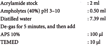
1. Prepare the gel mix as follows –
(for mini slab gel, 10 ml)
2. Pour the gel mix in the gel cassette and polymerize the gel. Then soak it in 10% Triton X-100 for 30 minutes.
(Including the Triton in the gel mixture interferes with adherence of the gel to the hydrophilic backing and so the polymerized gel is soaked later in Triton X-100.)
3. Perform IEF as follows –
i. Fix the gel in the electrophoretic apparatus.
ii. Fill the upper buffer chamber with 200 ml of cathode buffer and the lower buffer chamber with 200-400 ml anode buffer.
iii. Suspend the protein sample in IEF sample buffer and load.
iv. Run the gel for 60 minutes at 100 volts.
v. Run for 60 minutes at 200 volts.
vi. Then run for 30 minutes at 500 volts.
(Step-up increase in voltage prevents overheating and dehydration of the gel.)
4. Fix the gel for 15 minutes in the following fixing solution.
Sulfosalicylic acid (4%) – 10 g
Trichloroacetic acid (12.5%) – 31.25 ml of 100%
Menthol (30%) – 75 ml
Distilled water – to 250 ml
5. Stain the gel for 30minutes in –
Ethanol (27%) – 135 ml
Acetic acid (10%) – 50 ml
Distilled water – 15 ml
Coomassie R250 (0.04%) – 0.2 g
(Or)
CuSO4 (0.5 %) – 2.5 g
Distilled water – 500 ml
(Cupric sulfate helps to reduce the background staining of ampholytes; dissolve it in water before adding the alcohol.)
6. De-stain the gel for 30 minutes.
Ethanol (12%) – 60 ml
Acetic acid (7%) – 35 ml
Distilled water – 405 ml
7. Include IEF standards as pI markers.
Polyacrylamide (Stock):
Acrylamide – 24 g
Bisacrylamide – 1 g
Dissolve in 100 ml of distilled water.
Ammonium Persulfate (APS):
Dissolve 100 mg in 1 ml distilled water.
TEMED:
This is available as a commercial preparation.
Triton X-100 (10%):
Dissolve 1 ml in 10 ml distilled water (heat to dissolve).
These are available as commercial preparations in various pH ranges depending on the pI of the protein. If there is no idea about the nature of pI then pH 3-10 range is good to apply.
Triton X-100 (3%) – 300 µl of 10% stock
Ampholytes (2%) – 50 µl of 40% stock
DTT (20mM) – 20 µl of 1 M stock
Distilled water – 630 µl
A pinch of bromophenol blue
Anode Buffer (Anolyte):
0.5 M acetic acid – 200 ml
Cathode Buffer (Catholyte):
1.0 N NaOH – 500 ml
Method and Technique # 7. Trypsin Digestion of Protein Gel:
The choice of the fragmentation protein can be very important. If too many peptides are generated, spot information possibly will be lost. But if too few peptides are produced, they may be more difficult to isolate and analyze. In this method, selected gel spots are excised and enzymatically digested, and the tryptic peptides are purified for further analysis.
1. Dehydrate the gel spots for 10 minutes in 50 µl of 2:1 ratio of 50% acetonitrile and 25 mM ammonium bicarbonate for 15 minutes.
2. Remove the supernatant and rehydrate the spots for 10 minutes by adding 50 µl of 15 mM ammonium bicarbonate.
3. Remove the supernatant once again and dehydrate the spots.
4. Dry the spots in a speed vacuum for 1 hour.
5. Add 25 µl of 10 mM dithiothreitol in 25 mM ammonium bicarbonate for 1 hour at 56°C.
6. Remove the dithiothreitol and replace with 25 µl of iodoacetamide in 25 mM ammonium bicarbonate.
7. Incubate the spots for 45 minutes at room temperature in the dark.
8. Remove the supernatant and wash the spots with 25 µl of 25 mM ammonium bicarbonate for 10 minutes.
9. Dehydrate the spots with 50 µl of 2:1 ratio of acetonitrile and 25 mM ammonium bicarbonate for 15 minutes.
10. Rehydrate again and dehydrate, each for 10 minutes.
11. Dry the spots in speed vacuum for 1 hour.
12. Re-suspend the dried spots in trypsin (12.5 ng/µl) in 25 mM ammonium bicarbonate on ice. Hydrate the spots fully and incubate at 37°C for 4 hours.
13. Analyze these spots using PMF/MALDI-MS TOF.
Method and Technique # 8. Protein Dialysis:
The method offers considerations and comprehensive procedures for the use of dialysis to alter the composition of a protein solution. It is a useful step in adjusting a protein sample from one buffer to another and in correcting the metal- and salt-ion concentrations. It also helps in the removal of unwanted small molecules.
Dialysis tubing is basically a cylindrical membrane containing pores. The size of these pores determines the molecular weight ‘cut-off’ of the dialysis tubing. Typical cut-offs are 5 kD, 10 kD, 30 kD and 100 kD.
Thus, in tubing with a cut-off of 30 kD, all molecules smaller than 30 kD will be able to pass freely through the membrane. Dialysis tubing is available in dry and wet forms (as a ‘dry’ roll in a box whereas the other is wet and stored in a liquid buffer).
The dry roll must be pre-treated before use. This involves soaking the membrane in double distilled water, heating the membrane to 60°C in bicarbonate solution, and rinsing in double distilled water or tap water extensively. The wet membrane has already been pre-treated for use and only needs to be rinsed in double distilled water to remove preservatives.
1. After washing the dialysis tubing, calculate the length of tubing needed to contain the volume of protein sample. Cut the tubing to the required length, leaving an extra inch or two on each side for closures.
2. Use dialysis tubing-specific closure to close one end, or tie two tight knots on one end of the tubing.
3. Pipette the protein sample into the tubing and close the other end of the tubing with another closure or with two tight knots. Try to avoid having too many air bubbles inside the tubing.
4. Insert the dialysis tubing containing the protein into dialysis buffer taken in a large beaker. The volume of the buffer should be at least 100 times the original volume of the protein sample. The buffer should also be pre-chilled if the protein is labile.
5. Stir the buffer slowly with a stir bar on a magnetic stir plate for at least a few hours.
6. Discard the dialysis buffer and replace with the same volume of fresh dialysis buffer.
7. Dialyze for a few hours or overnight.
8. Remove the tubing from the beaker containing dialysis buffer and carefully open one end of the tubing. Pipette the dialyzed protein solution into tubes.
NaH2CO3 (2%)
EDTA (1 mM)
Metal and salt ions (NaCl, KC1, MgCl2)
Other small molecules (glycerol, DTT, ATP)
Typical protein buffering agents (Tris, HEPES, Phosphate-based)
Sometimes distilled water is also used as a dialysis solution.
Method and Technique # 9. Enzyme (Esterase) Gel Electrophoresis:
A non-denatured discontinuous gel electrophoresis system (Native PAGE) consisting of 7.5% resolving gel and 3% stacking gel is useful for most enzyme analysis. In this method, the electrophoresis of esterase enzymes is explained.
1. Grind 50 to 100 mg tissue with 200 ml of extraction buffer in a glass homogenizer.
2. Centrifuge the enzyme homogenate at 4°C for 20 minutes at 10,000 rpm.
3. Preserve the supernatant.
1. Mix resolving gel solution, gel buffer, ammonium persulfate (APS) and TEMED (see preparation of resolving gel) without forming any air bubbles.
2. Add this mixture into the gel mould (prepare as described for SDS-PAGE without using SDS), leave a gap of 1-2 cm for the stacking gel.
3. Add a layer of water saturated butanol on top of the gel to ensure an even surface while the gel polymerizes.
4. Prepare stacking gel using stacking gel solution, gel buffer, APS and TEMED (see preparation of stacking gel) without forming any air bubbles.
5. Pour off the butanol from the top of the polymerized resolving gel and rinse once with ethanol.
6. Pour the stacking gel on the top of the resolving gel. Carefully place the comb on the top of the gel mould to avoid trapping of air bubbles between the comb and gel.
7. Pre-run the gel with electrode buffer after fixing the gel sandwich to the electrophoresis apparatus for 30 minutes at 150 volts (for regular gel) and 80 volts (for mini gel).
8. Stop the pre-run. Mix the enzyme extract (10 µl) with marker dye (20 µl) and load the samples into the wells.
9. Run the gel for 5 minutes at 200 volts (for regular gel) and 120 volts (for mini gel) until the marker dye has clearly entered the gel.
10. Remove the gel sandwich and flush the wells with electrode buffer and restart the electrophoresis at 150 volts (for regular gel) and 80 volts (for mini gel) and continue running until the visible marker is three-fourths of the way down the gel.
11. Stain the gel with staining solution until the desired intensity of enzyme bands is apparent in the gel.
With this method very intensely stained red/purple or purple/blue bands should appear on the gel within minutes of esterase being elevated. 1-Naphthyl acetate specific bands are more blue coloured than 2-naphthyl acetate specific bands which are pink/red coloured.
12. Fix the gels in 10% acetic acid.
13. Calculate the relative mobility of enzyme using the following formula –
Rm = distance of enzyme migration/length of gel after staining x length of gel before staining/distance of dye migration.


