Humans suffer from several genetic disorders, which arise in the following ways:
1. Genetic Disorders Because of Change in Number of Autosomes:
a. Down’s Syndrome:
Down syndrome occurs because of trisomy 21. This takes place because of nondisjunction during oogenesis. This abnormality occurs in greater incidence in women above 40 years of age. The individual who suffers from Down’s syndrome has 47 chromosomes instead of 46 chromosomes.
It was first reported in the year 1866 by Langdon Down and is also called Mongolian idiocy or Mongolism. The syndrome is characterised by rounded face, broad forehead, flattened nasal bridge, open mouth, protruding tongue, short neck flat hands. The individual is mentally retarded and various organs are deformed.
The reproductive organs are under developed though a few victims attain maturity. Down’s disease may also arise by translocation of a segment or whole chromosome to the 14/15th chromosome. In such cases the total number of chromosomes remains intact. When the syndrome runs in the family it is known as familial Down’s syndrome.
Other examples of trisomy are as follows:
i. Edward’s Syndrome – trisomy of 18th chromosome
ii. Patau’s Syndrome – trisomy of 13th chromosome
b. Alzheimer’s Disease:
This disease is characterised by the progressive mental deterioration with loss of memory. The disease begins in the late middle age and leads to death in 5-10 years. It is caused by the accumulation of the amyloid protein in the brain leading to degeneration of the neurons. It is believed to be because of genes located in chromosomes on chromosome 21.
2. Genetic Disorders because of Change in Number of Sex Chromosomes:
Abnormalities in number of sex chromosomes occur during oogenesis or spermatogenesis because of non-disjunction. It results in the production of two types of eggs, i.e. XX and O eggs or two types of sperms, i.e. XY sperms and O sperm. Fertilisation of an abnormal gamete by a normal gamete results in the production of genetic disorders in the affected individuals.
The common disorders are described below:
a. Klinefelter’s Syndrome:
Klinefelter’s syndrome is characterised by XXY genotype (2n = 47 or 44 + XXY). It occurs from the union of a XX egg and a normal Y sperm or abnormal egg with a XY sperm. The affected individual is a sterile male and is characterised by unusually long body, obesity, with female characteristics such as breasts.
b. Turner’s Syndrome:
Turner’s syndrome occurs with a XO genotype (2n = 45 or 44 + X). It occurs from the union of an abnormal O egg with a normal sperm or a normal X egg with an abnormal O sperm. The individual has 45 chromosomes (2n-1). Turner’s syndrome is the only known viable monopsony in man. The affected individual is a sterile female with underdeveloped breasts, reduced ovaries, short stature, and with many male characteristics such as heavy neck muscles. She does not menstruate or ovulate.
3. Disorders Due to Mutation in Autosomes:
Mutations in the genes present in the autosomes can cause a number of genetic disorders in both males and females. They are called autosomal disorders. These disorders may be of two types – recessively inherited traits and dominantly inherited traits.
a. Recessively Inherited Traits:
These are caused by recessive genes expressed in homozygous condition.
The following are some of the examples of this type:
i. Alkaptonuria:
Alkaptonuria is a disease characterised by the blackening of urine on exposure to air. Urine of affected individuals contains homogentisic acid or alkapton which combines with oxygen to form the black pigment. In individuals with the normal gene, the homogentisic acid is broken down by homogentisate oxidase to compounds which enter the Krebs’s cycle to carbon dioxide and water. In affected individuals the enzyme is absent because of the abnormal gene (Fig. 21).
ii. Phenylketonuria:
Phenylketonuria occurs because of a recessive gene and is characterised by the absence of phenylalanine hydroxlase which is required for the conversion of amino acid, phenylalanine to another amino acid, tyrosine. In affected individuals phenylalanine accumulates in the tissues and some of it changes to phenyl pyruvic acid, which gets excreted in the urine. Accumulation of phenylalanine causes damage in the brain. The affected individual is affected from about six months of birth and is mentally retarded.
iii. Sickle Cell Anaemia:
This disease is because an autosomal recessive gene which affects the structure of haemoglobin.
iv. Albinism:
Albinism is also a recessively inherited disease which is manifested in a homozygous condition. It is characterised by the absence of the pigment, melanin in the skin, hair and iris. The affected individual is unable to see bright light. The disease is caused by the absence of an enzyme known as tyrosinase which is necessary for the synthesis of melanin from dihydroxy-phenylalanine.
b. Dominantly Inherited Traits:
The dominantly inherited traits are caused by dominant genes which expresses itself in either homozygous or heterozygous condition. Even if one parent carries the defective gene, half the children will inherit it (Fig. 22).
Some examples of dominantly inherited traits are listed below:
i. Brachydactyly:
A condition where affected individuals have abnormally short digits.
ii. Polydactyly:
A condition where the affected individuals have extra digits.
iii. A disorder where the crown of the teeth wear off quickly.
iv. A form of dwarfism.
v. Huntington’s Chorea:
This is a dominant autosomal disorder due to an allele on the short arm of chromosome 4. The disease manifest at the age of 15-40 years or sometimes later. The brain atrophies resulting in faulty speech and irregular movements of the limbs.
4. Disorders Due to Mutation in Sex Chromosomes:
Many disorders arise because of mutation in the genes present on the sex chromosomes. These traits are known as sex linked disorders and the transmission of these is known as sex linked inheritance. The recessive gene on the X chromosome affects the males more than the females.
This is because males suffer the disorder when the mutated gene is present in a single dose since he possesses only a single X chromosome, while females suffer the disorder only on homozygous condition. When a female possesses a single gene in heterozygous condition, she is known as a carrier.
Some sex linked disorders are described below:
a. Haemophilia:
Haemophilia is also known as the bleeder’s disease reported in 1803. The affected individual is unable to stop bleeding in case of minor skin cuts and may bleed to death. This is because of the absence of factor VIII, a protein required for clotting of blood.
The normal gene which is responsible for the production of the protein required for clotting of blood is located on the X chromosome. The mutated gene can be expressed as Xh. The inheritance of haemophilia is shown in Fig. 23. The inheritance of sex linked inheritance is also known as criss-cross inheritance.
b. Daltinism or Red Green Colour Blindness:
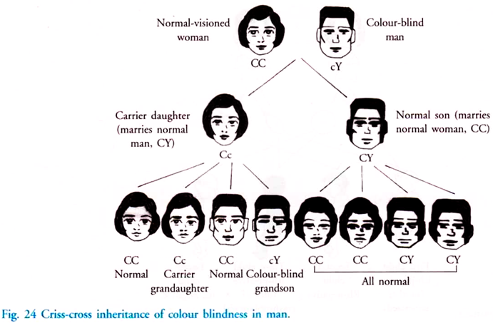
In Daltinism the affected individual is unable to distinguish between red and green and is caused by X linked recessive gene. The inheritance of daltinism is similar to haemophilia. The mutated gene is indicated as Xc (Fig. 23).
c. Night Blindness:
Night blindness is caused by a recessive gene located on the X chromosome. This causes a deficiency in the functioning of the retinal rods. This condition is also known as congenital night blindness. It can also occur in individuals because of vitamin A deficiency. This condition is known as acquired night blindness. The inheritance of the congenital night blindness is similar to above mentioned examples.
d. Muscular Dystrophy:
Muscular dystrophy is characterised by the deterioration of muscles at an early age. Because of the mutation, the body does not produce a protein known as dystrophin. Dystrophin is required to release calcium from the storage areas in muscle cells during muscular contraction. When the protein is absent, calcium is not released and muscle contraction does not occur. The affected individual becomes an invalid at the age of 10 and dies by the age of 20.
5. Disorders Due to Incompatibility of Genes:
Genes express themselves in the body by directing synthesis of chemical substances. When the chemical substances of male and female are not compatible with each other, the offspring born to these two individuals become affected. The incompatible chemical substances will harm the offspring, and in some cases prove fatal. The chemicals which produce this condition include the Rh factor and the other antigen-antibodies present in the blood.
a. ABO Incompatibility:
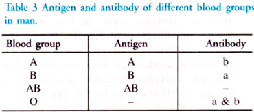
Blood groups in man are of four types based on the antigen and antibodies they possess. When two different groups of blood are mixed, the red blood corpuscles clump. This process is known as agglutination. Clumping occurs when the antigens present on the RBC of one individual combine with the antibodies present in the plasma of the other individual.
The incompatibility of blood groups is expressed during blood transfusion and during pregnancy. During blood transfusion, the antigen in the donor must be compatible with the antibody of the recipient. If they are incompatible the result may prove fatal.
b. Rh Incompatibility:
Rh factor is a protein on the surface of the red blood corpuscle. About 95% of the population possess this factor and are categorized as Rh positive. The formation of this factor is controlled by a dominant gene designated as ‘R’. The remaining 5%, who do not possess this factor, are called Rh negative. The recessive gene in Rh negative individuals is designated as ‘r’. Both the Rh positive and Rh negative individuals are normal in all respects. The problem arises during blood transfusion and during pregnancy.
When a Rh negative individual receives blood from a Rh positive individual for the first time, no harm is caused. But Rh negative individual starts producing anti-Rh factors or antibodies in response to the Rh factor of the donor. If the same individual receives blood for the second time from a Rh positive individual, the anti-Rh factors react with the Rh antigen of the donor and the recipient may die.
The Rh factor also has great significance during childbirth. If a Rh negative woman is sensitised by Rh positive blood, the children born to the woman may be affected. This can also happen if she gets married to a man as shown in Fig. 24. The children may inherit the Rh antigen from the father.
In such cases, the antibodies from the mother may pass through the placenta and damage the red cells of the child during the last months of pregnancy. This causes the disease known as erythroblastosis foetalis. During this condition, the child suffers from anaemia due to haemolysis or breakdown of the red blood cells in the foetus and consequent jaundice as the blood vessels in the liver becomes blocked with broken cells and the bile is absorbed into the blood.
The disease may be severe so as to cause death before birth (still birth) or after birth (neonatal birth). This is also known as haemolytic disease Normally the first child will not be affected since the antibodies do not reach sufficient strength to harm the baby. Subsequent positive children may become affected.


